Lower Leg Bones Diagram : Bone Fracture_Human Leg Anatomy And Skeleton Stock Vector - Illustration of foot, lower: 114048504. Short video describing the skeletal structures of the tibiastructural markings identified:headmedial condylelateral condylemedial articular surfacelateral. This diagram depicts lower leg bones 1024×1350. Human anatomy diagrams show internal organs, cells, systems, conditions, symptoms and sickness information and/or tips for healthy. The fibula is a long, skinny lower leg bone that looks rather fragile. Anterior view with primary bones names.
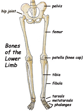
Master leg and knee anatomy using our topic page. Your legs are two of your most important body parts. Leg length discrepancy (lld) has been a controversial issue among researchers and clinicians for many years. While their parts are similar in general, their structure has been adapted to differing functions. The bones of the leg are the femur, tibia, fibula and patella.
18 Best Images of Leg Anatomy Worksheets - Lower Limb Muscles Anatomy Worksheet, Lower Limb ... from www.worksheeto.com Two bones make up the lower arm. Continue scrolling to read more below. Diagram of lower leg bones posted on march 25, 2019 by admin this image shows the structure of tibia and fibula left panel legs bone diagram 20 13 asyaunited de u2022 hip drawing outline foot overview of bones the lower limb posterior and anterior view respectively 62 infographic diagram of. However, in the world of anatomy, the 'leg' strictly means. Its presence is accepted but. Anterior view with primary bones names. Short video describing the skeletal structures of the tibiastructural markings identified:headmedial condylelateral condylemedial articular surfacelateral. The tibia (shin bone) is the medial bone of the leg and is larger than the fibula, with which it is paired (figure 3).
Anterior view with primary bones names.
Good front and back human body skeleton diagram with bones identified. The human leg, in the general word sense, is the entire lower limb of the human body, including the foot, thigh and even the hip or gluteal region. Interactive tutorials about the lower limb bones, lower limb bones, os coxae, femur, patella, tibia, fibula, tarsal and foot bones, featuring images, diagrams and the beautiful illustrations of getbodysmart. The tarsals are ankle bones and, along with the other bones in the foot (the metatarsals and phalanges), support weight and act as shock absorbers for the body. Your upper and lower leg are connected by a hinge joint. Bone contusions, osteonecrosis, marrow edema syndromes, and stress > fractures) > infections of bone, joint, or soft tissue (eg. Leg bones, hip bone, back bone, etc.) what do we call a person who has lost a body part? However, the definition in human anatomy refers only to the section of the lower limb extending from the knee to the ankle, also known as the crus. Anterior view with primary bones names. Two bones make up the lower arm. Lower jaw (mandible) collar bone. Leg length discrepancy (lld) has been a controversial issue among researchers and clinicians for many years. Continue scrolling to read more below.
While their parts are similar in general, their structure has been adapted to differing functions. The foot bones shown in this diagram are the talus, navicular, cuneiform, cuboid, metatarsals and calcaneus. Cheek bone (zygoma) upper jaw (maxilla). Bone contusions, osteonecrosis, marrow edema syndromes, and stress > fractures) > infections of bone, joint, or soft tissue (eg. Two bones make up the lower arm.
Aluminium Plant Safety: "cited after worker's leg amputated...." from 3.bp.blogspot.com Leg length discrepancy (lld) has been a controversial issue among researchers and clinicians for many years. Your legs are two of your most important body parts. Leg bones, hip bone, back bone, etc.) what do we call a person who has lost a body part? Your leg bones are the longest and strongest bones in your body. The tibia (shinbone) is the large bone of the lower leg, and the fibula is the thin bone along the outer aspect of the lower leg. When you stand or walk, all the weight of your upper body rests on them. Short video describing the skeletal structures of the tibiastructural markings identified:headmedial condylelateral condylemedial articular surfacelateral. Continue scrolling to read more below.
The foot bones shown in this diagram are the talus, navicular, cuneiform, cuboid, metatarsals and calcaneus.
Its presence is accepted but. Your upper and lower leg are connected by a hinge joint. Continue scrolling to read more below. They allow you to move and provide support for your upper body. Good front and back human body skeleton diagram with bones identified. At the microscopic level, this hard outer shell is made up of rod like structures called osteons. The fibula is a long, skinny lower leg bone that looks rather fragile. The musculoskeletal segment of the leg, including the foot bones (ankle, heel bone, toe bones), fibula and tibia, knee, femur and. At the distal end of the femur, two rounded condyles meet the tibia and fibula bones of the lower leg to form the knee joint. Your legs are two of your most important body parts. Lower jaw (mandible) collar bone. The tarsals are ankle bones and, along with the other bones in the foot (the metatarsals and phalanges), support weight and act as shock absorbers for the body. Two bones make up the lower arm.
When you stand or walk, all the weight of your upper body rests on them. The radius is along the thumb side and the ulna is (answers: We'll break down the anatomy and function of the upper leg, knee, lower leg, ankle, and foot. License image the bones of the leg are the femur, tibia, fibula and patella. Anterior view with primary bones names.
Skeletal Series Part 10: The Human Leg | These Bones Of Mine from thesebonesofmine.files.wordpress.com The two bones beneath your knee that make up your shin are your tibia and fibula. The tibia (shin bone) is the medial bone of the leg and is larger than the fibula, with which it is paired (figure 3). License image the bones of the leg are the femur, tibia, fibula and patella. Lower jaw (mandible) collar bone. Leg length discrepancy (lld) has been a controversial issue among researchers and clinicians for many years. Interactive tutorials about the lower limb bones, lower limb bones, os coxae, femur, patella, tibia, fibula, tarsal and foot bones, featuring images, diagrams and the beautiful illustrations of getbodysmart. Most bones (particularly the long bones of the arms and legs — which make up the appendicular skeleton) have a hard outer shell known as cortical bone. Its presence is accepted but.
Your leg bones are the longest and strongest bones in your body.
The fibula is a long, skinny lower leg bone that looks rather fragile. Continue scrolling to read more below. Your legs are two of your most important body parts. Depending on the cause, leg lumps may be single or while these conditions are rare, both benign and malignant tumors of the skin, soft tissues, or bones can sometimes feel like leg lumps. Physical performance conflict, for example, difficulties walking or climbing stairs, not being able to keep up, a poor performance leg segment: The tibia (shin bone) is the medial bone of the leg and is larger than the fibula, with which it is paired (figure 3). Lower jaw (mandible) collar bone. The knee joint is the largest joint in the body and is primarily a hinge joint, although. Bone contusions, osteonecrosis, marrow edema syndromes, and stress > fractures) > infections of bone, joint, or soft tissue (eg. Your upper and lower leg are connected by a hinge joint. The knee joint is the largest joint in the body and is primarily a hinge joint, although some sliding and rotation occur. They allow you to move and provide support for your upper body. At the distal end of the femur, two rounded condyles meet the tibia and fibula bones of the lower leg to form the knee joint.
Interactive tutorials about the lower limb bones, lower limb bones, os coxae, femur, patella, tibia, fibula, tarsal and foot bones, featuring images, diagrams and the beautiful illustrations of getbodysmart leg bones diagram. At the distal end of the femur, two rounded condyles meet the tibia and fibula bones of the lower leg to form the knee joint.
Share :
Post a Comment
for "Lower Leg Bones Diagram : Bone Fracture_Human Leg Anatomy And Skeleton Stock Vector - Illustration of foot, lower: 114048504"


Post a Comment for "Lower Leg Bones Diagram : Bone Fracture_Human Leg Anatomy And Skeleton Stock Vector - Illustration of foot, lower: 114048504"